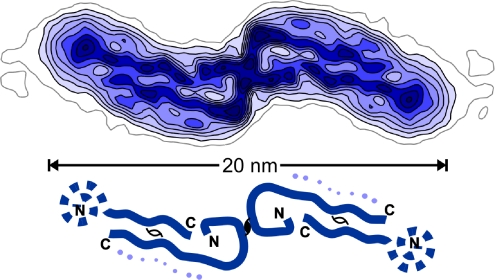
Fig. 1.
Reconstructed cross-section density map at ≈8-Å resolution of the double protofilament “20-nm” Aβ(1-40) fibril (1, 3). The schematic diagram indicates the likely contour of the quasi-symmetrically paired β-strands identified in each protofilament, one traced along its 40-residue length and the other with a disordered tail. Faint density along side the long arm of the more ordered β-strand, indicated by dots in the diagram, may represent the fractional molecule per 4.7-Å repeat whose structure is very variable. N and C termini were identified in ref. 3. This figure was prepared by M. Schmidt (Brandeis University, Waltham, MA).

