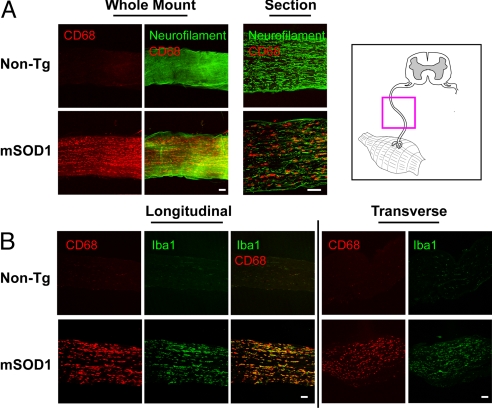
Fig. 2.
Sciatic nerves in mutant SOD1 mice but not WT mice show intra-axonal activation of macrophages. (A) Whole-mount segments and longitudinal sections of distal sciatic nerve from end-stage mSOD1G93A and non-Tg litter-mates were stained for macrophages (CD68) and axons (neurofilament). For whole-mount stains, confocal microscopy images 100 μm into the nerve are shown as a composite. (B) Longitudinal and transverse sciatic nerve sections were stained for macrophage markers CD68 (red) and Iba1 (green). mSOD1G93A nerves at end stage show an abundance of activated, CD68/Iba1+ macrophages compared with non-Tg litter-mates. An anatomical schematic of peripheral nerves is shown (Upper Right). (Scale bars: 100 μm.)

