Fig. 1.
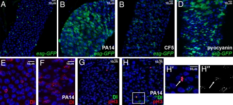
P. aeruginosa infection induces intestinal progenitor expansion. (A–D) Posterior midgut cells of uninfected (A), strain PA14 infected (B), strain CF5 infected (C) and pyocyanin-fed (D) flies. SCs and progenitors of esg-GAL4 UAS-srcGFP flies fed for 5 days are shown in green. (E and F) Posterior midgut SCs marked with α-Delta antibody (red) of uninfected (E) and PA14-fed (F) flies. (G, H) Posterior midgut SCs marked with α-Delta (green) and phospho-histone-H3 (pH3; red) antibody, of uninfected (G) and PA14-fed (H) flies. (H′ and H″) Magnification of rectangular region in (H), showing colocalization of pH3 (H′; arrow) and Delta (H′ and H″; arrow) staining. Nuclei in all panels are marked with DAPI (blue).

