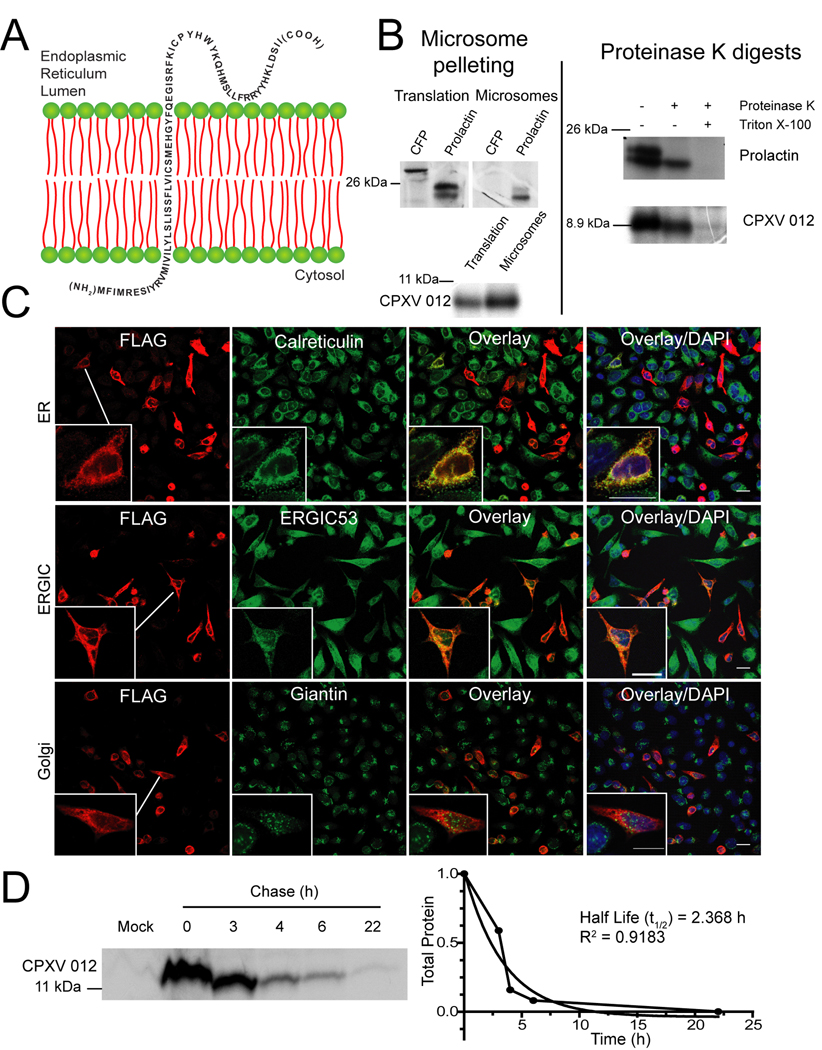
Figure 2. CPXV012 is a short lived, ER resident type II transmembrane protein.
(A) Model of CPXV012 topology in the ER membrane. (B) In vitro translation of CPXV012 mRNA in the presence of microsomes. In vitro transcribed mRNAs encoding CPXV012 and control proteins, CFP and prolactin were added to a rabbit reticulocyte translation system in the presence of canine microsomal membranes. Left panel: Total translation products or products recovered after microsome pelleting. Right panel: Following translation, pelleted microsomal fractions were treated with proteinase K and separated by SDS-PAGE. Molecular weight markers (kDa) are shown. (C) Subcellular localization of CPXV012. HeLa cells were transiently transfected with pCPXV012-N-FLAG. After 48 h, cells were fixed, permeabilized, and stained with antibodies specific to calreticulin, ERGIC53, giantin, or FLAG. (D) CPXV012 half-life by pulse-chase. pCPXV012-CON-FLAG and empty vector control (Mock) were transiently transfected into HeLa cells. At 24 hpt, cells were pulse-labeled for 1 h and the label was chased for the indicated time. Lysates were immunoprecipitated with anti-FLAG, separated by SDS-PAGE, and visualized by autoradiography (left panel). Optical density of CPXV012-specific bands was measured and plotted against the chase time (right panel).

