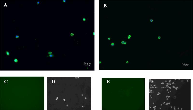
Figure 5.
Epifluorescence imaging of cells labeled with the trivalent construct (0.625 μM) incubated at 37°C followed by trypsin treatment. After treatment with trypsin, NHA (panel A) and SF 767 cells (panel B) demonstrate persistent fluorescence indicating internalization of the construct. Panels C through E (C & D correspond NHA while E & F correspond to SF 767) depict epifluorescence and phase imaging of cells treated with the trivalent construct (0.625 μM) incubated at 4°C followed by trypsin treatment. No binding is visible on the cells after trypsin treatment.

