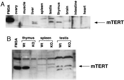
Figure 5.
Expression of mTERT protein in different murine tissues. (A) Western blot analysis of S-100 extracts from the indicated tissues, using K-370 antibodies. Tissues were obtained from wild-type mice. One hundred micrograms of total protein was loaded per lane. FM3A is a mouse cell line expressing high levels of mTERT (see Fig. 4). The distortion in the lane corresponding to liver is probably because of the presence of an abundant protein in the relevant region of the gel. Band specificity was confirmed by preincubation of the K-370 antibodies with the antigenic peptide (not shown). (B) Western blot analysis of S-100 extracts of thymus, spleen, and testis obtained from wild-type and mTR−/− littermates, and using K-370 antibodies. One hundred micrograms of total protein was loaded per lane, except in the case of the FM3A extract, of which only 20 μg was loaded. The specificity of the mTERT band was determined by preincubation of the K-370 antibody with the antigenic peptide (not shown).

