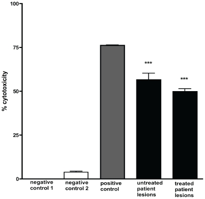
Figure 4. Cytotoxicity of ASL from human Mu lesions on human embryonic lung fibroblasts.
Cytotoxicity after 48 h culture was assessed using an MTT assay. Negative control 1 is untreated cells, negative control 2 is ASL from uninfected skin. Positive control was purified mycolactone at a concentration of 5 µg/ml. Significant cytotoxicity was observed with all patient samples with ***p<0.001 compared to negative control 1. The apparent difference in percentage cytotoxicity between 5 untreated and 5 antibiotic treated lesions was not statistically significant. HELF cells were treated in quadruplicates and cytotoxicity determined in at least 2 independent experiments. Data are shown as a percentage of untreated cells (negative control 1). Error bars are ±SEM of duplicate assays.

