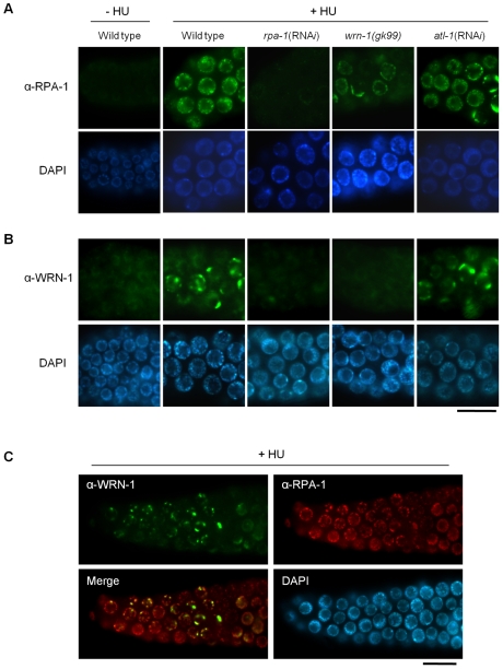
Figure 4. Reciprocal dependence of RPA-1 focus formation and nuclear localization of WRN-1 after DNA replication inhibition.
One-day old adult wild-type N2, rpa-1(RNAi), wrn-1(gk99), and atl-1(RNAi) worms were treated with hydroxyurea (25 mM) for 8 h. Knockdown of atl-1 and rpa-1 was carried out as in Figure 2 but from the L4 stage for 16 h, and premeiotic germ cells were stained with antibodies against (A) RPA-1 and (B) WRN-1. (C) Partial colocalization of RPA-1 and WRN-1 in the nuclei of premeiotic germ cells after hydroxyurea treatment. All the WRN-1 spots overlap with RPA-1 foci but not vice versa. Magnification bars, 10 μm.

