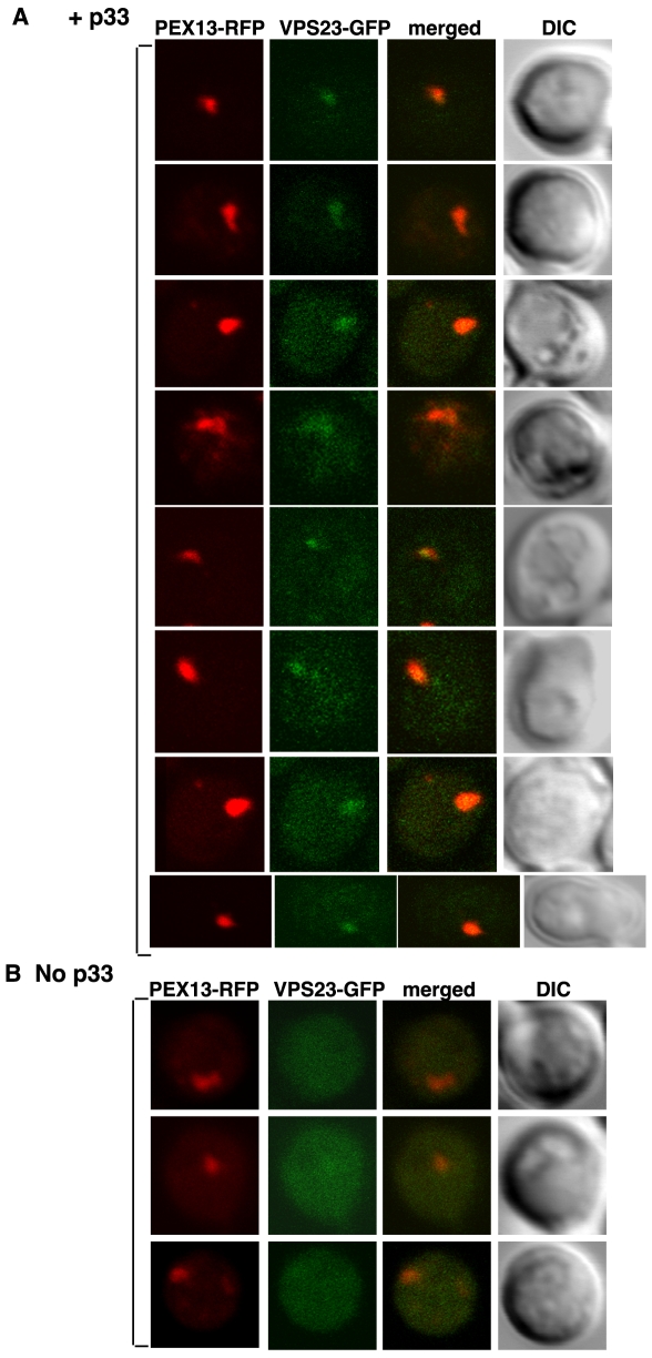
Figure 7. Partial re-distribution of Vps23p to the yeast peroxisomal membranes in the presence of p33.
(A) Confocal laser microscopy images show the subcellular localization of Vps23p-GFP in the presence of p33 expressed from CUP1 promoter for 15–45 minutes in yeast strain DKY79 (VPS23:GFP, vps4Δ; vps27Δ). The peroxisomes were visualized with Pex13p-RFP marker. The merged images show the co-localization of Vps23p-GFP and Pex13p-RFP marker. DIC (differential interference contrast) images are shown on the right. Each row represents a separate yeast cell. (B) Cytosolic localization of Vps23p-GFP in the absence of p33. Yeast was grown under similar conditions and images were taken as in panel A.

