
Figure 6. Double immunostaining of SDF-1α and CD31 in gastrocnemius and thigh muscle 3 days after acute vs. gradual occlusion.
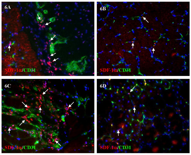
Arrows showed part of SDF-1α positive cells after acute (A) and gradual (B) occlusions in gastrocnemius. 6C and 6D showed the similar results of SDF-1α and CD31 double staining in thigh muscle after acute (6C) and gradual occlusion (6D).