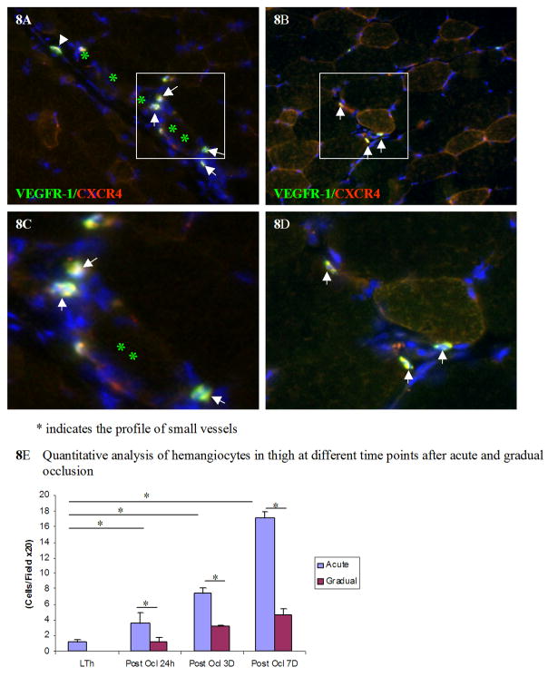
Figure 8. More hemangiocytes are integrated into the small vessels in thigh muscle 7 days after acute occlusion.
More hemangiocytes were detected in thigh muscle at post occlusion day 7 after acute occlusion (8A) than after gradual occlusion (8B). The enlargement of images 8A and 8B are shown in 8C and 8D respectively. Asterisks show the lumen of small vessels. Arrows show the hemangiocytes, which are double positive for both VEGFR-1 and CXCR4. Hemangiocyte line along the endothelium of a small vessel. The quantitative analysis of hemangiocytes in each field at different time points post occlusion is shown in Figure 8E. * indicates p<0.01

