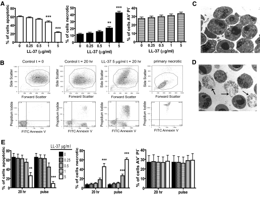
Figure 1.
Induction of PMN necrosis by LL-37. (A–D) Freshly isolated human PMN were incubated for 20 h over a range of LL-37 concentrations before analyses. Primary necrosis was induced by heating at 65°C for 30 min. Cell death was examined by FACS analyses and TEM morphology. (A) Apoptotic (FITC-AV-positive, PI-negative), necrotic (FITC-AV-positive, PI-positive), and live (FITC-AV-negative, PI-negative) cells were enumerated. Figures represent mean values ± sem for n = 28 different donors for each condition, performed in triplicate. Significance was assessed by one-way ANOVA with Bonferroni’s multiple comparison test comparing each treatment with control; **, P ≤ 0.01; ***, P ≤ 0.001. (B) Representative FACS plots. (C and D) Representative TEM images of (C) untreated, apoptotic PMN and (D) PMN exposed to 5 μg/ml LL-37. Arrows indicate examples of necrotic PMN. (E) Freshly isolated human PMN were incubated for 20 h over a range of LL-37 concentrations (20 h samples) or incubated for 20 h in the absence of LL-37 followed by exposure to a range of LL-37 concentrations for 10 min (pulse samples) before FACS analyses of apoptotic, necrotic, and live cells. Panels represent mean values ± sem for n = 3 donors performed in triplicate. Significance was assessed by two-way ANOVA with Bonferroni’s post-test; **, P ≤ 0.01; ***, P ≤ 0.001, compared with untreated control.

