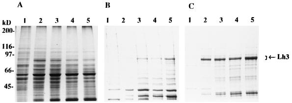
Figure 3.
Analysis of the expression of the human lysyl hydroxylase 3 polypeptide in insect cells by SDS/PAGE under reducing conditions. (A) NP-40 soluble proteins. (B) Glycerol buffer soluble proteins. (C) Proteins solubilized from the remaining pellets with 1% SDS. Lanes 1–5, samples from High Five cells infected with the virus coding for lysyl hydroxylase 3 and harvested 24, 40, 48, 64, and 72 hr after infection, respectively. Arrow indicates the location of the lysyl hydroxylase 3 polypeptides. Gels were stained with Coomassie brilliant blue.

