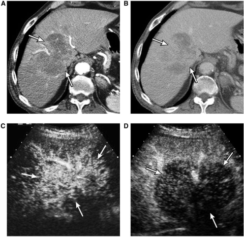
Figure 7.
An 82-year-old male with cholangiocarcinoma. (A) Arterial phase CT scan shows a large mass (arrows) with irregular peripheral enhancement. (B) Three-minute delay phase CT scan shows progression of enhancement within the mass (arrows). (C) Contrast-enhanced sonogram at 19 s delay shows hypervascularity of the mass (arrows). (D) Contrast-enhanced sonogram at 34 s delay shows early complete washout of enhancement of the mass (arrows), discordant with the CT scan (B).

