Fig. 3.
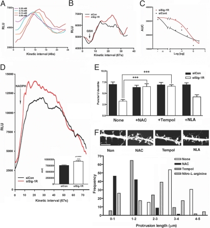
Free radicals in Sig-1R-knockdown neurons. (A) Generation of superoxide anions by GSH and sodium selenite. Sodium selenite was mixed with GSH at 0.2 mg/mL. O2·− were monitored by lucigenin-based chemiluminescense (lucigenin at 50 μM). (B) Effect of hippocampal neuronal lysates on O2·− generated by GSH/selenite. Neurons were transduced with AAV-siCons or AAV-siSig-1Rs for 10 days before assay. (C) The AUC, calculated from assays in panel B (between the 10th and 25th intervals), seen with increasing lysates (0, 0.03, 0.1, 0.3, 1, and 3 μg protein). (D) Effect of AAV-siSig-1R-td on the NADPH-induced generation of O2·− in living hippocampal neurons. NADPH at 45 μM was applied to neurons in the presence of 50 μM lucigenin at the beginning of the fifth interval (AUC from 10th to 40th interval). (E) Inhibition of AAV-siSig-1R-td-induced mitochondrial permeability transition by free radical scavengers NAC and Tempol. The puncta of JC-1 positive mitochondria in dendrites were measured. ***P < 0.001; n = 5, each group in triplicates. (F) Effects of free radical scavengers on dendritic spine formation in AAV-siSig-1R-td neurons. Neurons were transduced with AAV-siSig-1Rs on DIV 13 when 50 μM Tempol, NAC, or NAL were also added. Protrusion lengths were measured on day 5 posttransduction. Frequencies of various protrusion lengths are shown; n = 3 independent experiments. Representative morphologies are shown at the Top. (Scale bar, 5 μm.)

