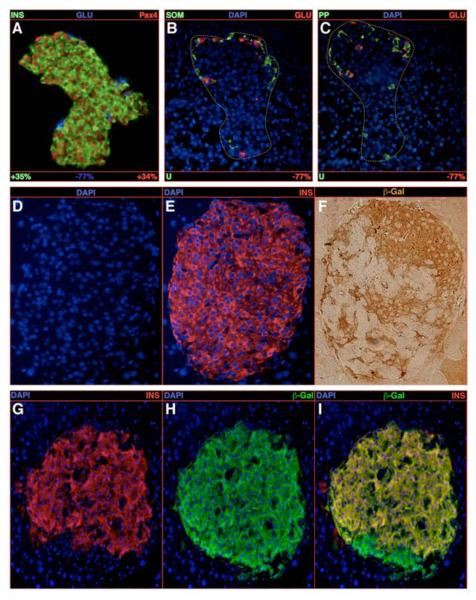
Figure 3. Conversion of glucagon-expressing cells into insulin-producing cells upon Pax4 ectopic expression.
(A-C) Quantification of the endocrine cell content alterations upon ectopic expression of the Pax4 gene in glucagon-producing cells in 1-week-old animals. A clear increase in the number of insulin-/Pax4-labeled cells at the expense of glucagon-expressing cells is highlighted (A), whereas the δ- and PP-cell contents are found unchanged (B-C). Note the accumulation of the remaining glucagon-marked cells at one pole of the islet. (D-F) The detection of insulin- and β-galactosidase-expressing cells on serial sections demonstrates that numerous insulin-labeled cells do express the β-galactosidase gene that normally marks glucagon-positive cells. (G-I) The same observation is made in 6-week old animals using co-immunofluorescence. All reported values are statistically significant (P<0.05, n=5). See Table S2 for a detailed analysis.

