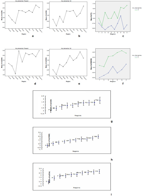
Figure 4.
Similarity of distributional trends of CD8, CD68 and P24 in the brain. Linear models were used to assess the effect of region within patients. We observed that the deeper midline and mesial temporal structures of the brain were much more involved than other CNS regions as evidenced by CD8 infiltration (a), CD68 infiltration (d) along with P24 antigen (i), respectively, in HAD patients. In contrast, in HIV non-dementia patients, very similar and comparable distributional trend was found at the level of CD8 (b) and CD68 (e) infiltration patterns, although there was complete absence of P24 antigen staining across all the regions. When the data of CD8 and CD68 of HAD and HIV non-dementia patients were combined (g and h), not surprisingly the trends are very similar with P24 distributional trend in HAD patients. The CTL and activated macrophage results confirm that trend again (c and f).

