
Figure 5.
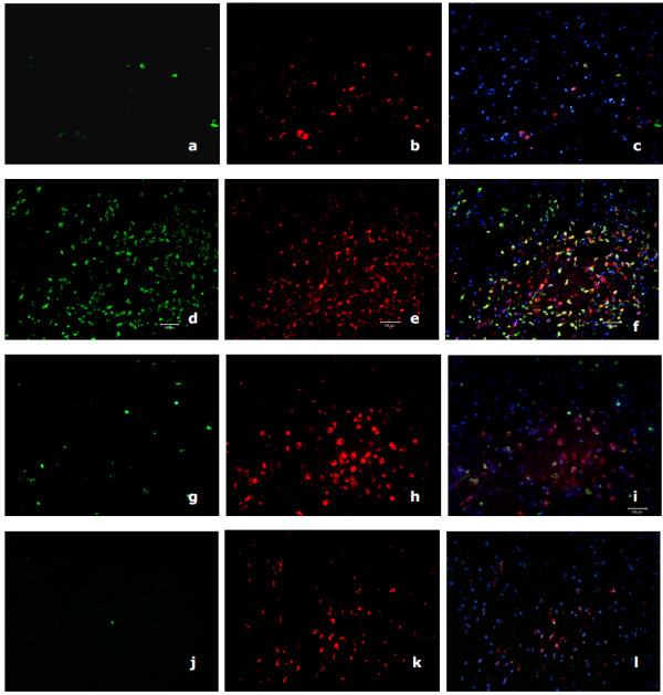
Immunohisto-fluorescent S100/CD68 double staining of HAD and HIV non-dementia patient. Immunohisto-fluorescent S100(green)/CD68(red) double staining of HAD (patient A parietal lobe: a, b and c; patient A medulla: d, e and f) and HIV non-dementia (patient D medulla: g, h and i; patient F medulla: j, k and l) patient. These data comply with our immuno-histochemistry staining in the deeper midline and mesial temporal structures of the brain. The CD68 staining was much more pronounced than lobes and there are comparable levels of CD68 staining, while the S100 stained cells are significantly less in HIV non-dementia patients.