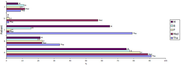
Figure 6.

Percentage of activated macrophages in the deeper midline and mesial temporal structures of the brain. Percentage of activated macrophages in the deeper midline and mesial temporal structures of the brain, based on the ratio of S100A8+CD68+ cells count to CD68+ cell counts in each of the CNS regions shown. In general, high activation rate was observed in HAD patients, except the mid brain and thalamus in the patient C and medulla in patient D. M: mid brain, B: basal ganglia, P: pons, MED: medulla, Tha: thalamus.
