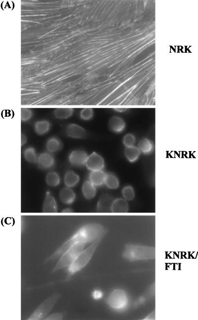
Figure 3.
Immunofluorescence microscopy of actin stress fibers. NRK and KNRK cells were seeded on 4-well chamber slides and treated the following day with DMSO or with 20 μM SCH56582. After 24 h, cells were washed and fixed in 4% paraformaldehyde and stained with fluorescein isothiocyanate-labeled phalloidin as described in Materials and Methods. (A) NRK. (B) KNRK. (C) KNRK treated with 20 μM SCH56582. The stained images were captured at ×400 magnification. At least three experiments were carried out with results similar to those shown here.

