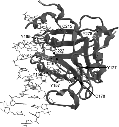
Fig. 1.
Crystal structure of AAG bound to εA-containing DNA. The structure was generated using the UCSF Chimera software package (23) and protein databank file 1F4R (24). Tyrosines and cysteines are represented by a gray ball and stick structure within the ribbon structure of AAG. The flipped εA is shown in gray as a stick structure.

