
Figure 5.
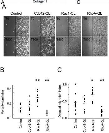
Migration of RhoA-QL, Rac1-QL, Cdc42-QL and control fibroblasts. Small colonies (10 µl) of Cdc42-QL, Rac1-QL, RhoA-QL and control cells were seeded in serum-containing medium in the center of tissue culture wells and allowed to attach at 37 °C. After 1 h, the cells were washed several times with serum-free medium and the wells were filled with serum-free medium. At this point, cell migration was recorded at the border of the colonies by time-lapse video microscopy for 800 min, with one picture every 10 min (supplemental movies). Pictures in A show the colony margin of control and QL cells at the onset (T0) and end of recording (T800). Single-cell tracking was used to determine the velocity (B) and directed migration index (C) of the different clones as described in Materials and methods. *p<0.02 and **p<0.005 versus mock-transfected (control) cells (Student’s t test; n = 20 for each group).