Figure 4.
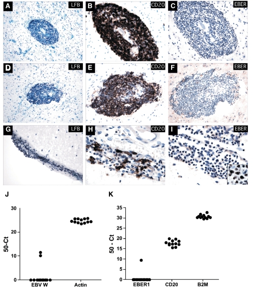
EBV was detected in very few multiple sclerosis specimens containing B cell infiltration within the meninges and parenchymal B cell aggregates. Fixed-frozen tissue specimens were sectioned with minimal waste and stained with LFB, CD20 and EBER. LFB staining identified B cell aggregates in regions of considerable demyelination (A and D—Multiple sclerosis_UK10) (blue = positive staining for myelin; pink = negative for myelin). Dense CD20+ B cell aggregates are shown in the brain parenchyma in one case (B and E—Multiple sclerosis_UK10), with loose B cell infiltrates identified within the meninges (G) of several others (H and not shown), while most cases had a diffuse B cell infiltrate. ISH revealed no EBER+ in all 12 multiple sclerosis specimens examined (C, F and I and not shown). An EBV-positive CNS lymphoma (I inset) served as a positive control for EBER ISH. Snap-frozen multiple sclerosis tissue specimens from the same cases were serially sectioned (10 μm sections; see Methods section) for alternate DNA or RNA isolation. Genomic EBV (J) was detected in 2 of 12 multiple sclerosis samples examined while EBER1 (K) was detected in one sample with detectable genomic EBV. All samples had a detectable CD20-positive B cell infiltrate (K). Note the sample with detectable genomic EBV but not EBER1 only contained EBV within one of two tissue sections examined (value reported is the average seen within the positive section only). Figures A and D (200×); B, C, E, F and G (400×); H, I and inset (1000×).

