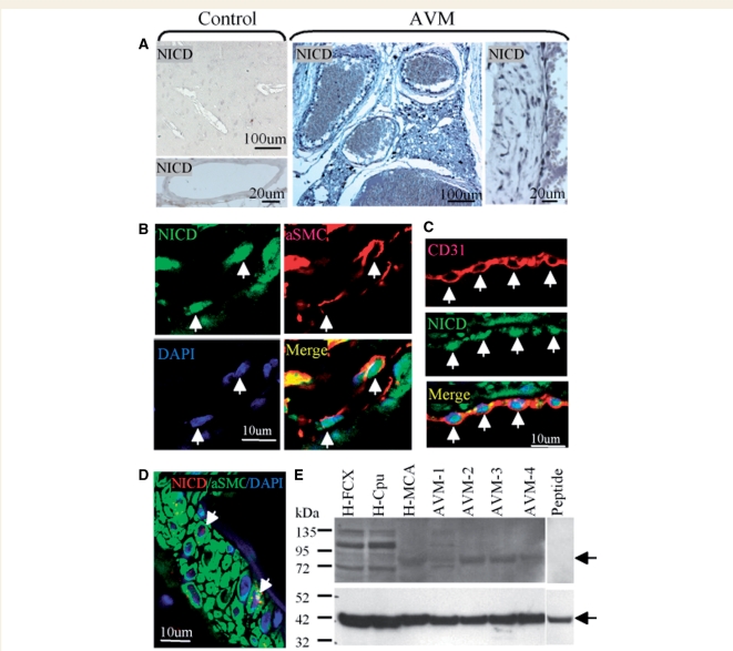
Figure 1.
Activation of Notch-1 signalling in human brain AVMs. (A) NICD was barely detectable in normal human cerebral vessels (Control), but was highly expressed in cell nuclei of AVMs. (B) Double immunostaining of human brain AVM shows nuclear NICD (green) and cytoplasmic SMC (red) in the same (vascular smooth muscle) cells (arrows). DAPI (blue) was used to counterstain nuclei. (C) Double immunostaining of human brain AVM shows nuclear NICD (green) and cytoplasmic CD31 (red) in the same (endothelial) cells (arrows). DAPI (blue) was used to counterstain nuclei. (D) Double immunostaining of normal human middle cerebral artery shows abundant cytoplasmic SMC (green) but little nuclear NICD (red). DAPI (blue) was used to counterstain nuclei. (E) Protein was isolated from normal human frontal cortex (H-FCX), caudate-putamen (H-Cpu) and middle cerebral artery (H-MCA) and from four human AVMs (AVM 1–4). Western blotting was performed using an antibody against NICD (top panel). A band of the predicted size (arrow) was blocked after pre-incubating anti-NICD with an excess of immunizing peptide (Peptide). The membrane was re-probed with anti-actin as an internal protein loading control (bottom panel, arrow).

