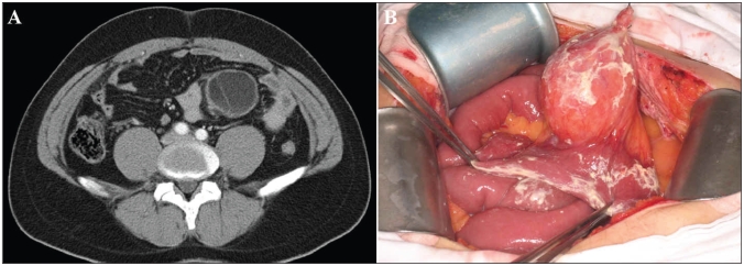
Fig. 1.
(A) Computed tomography scan of the abdomen of a 31-year-old man showing a homogenous tumour, 8 × 6 × 6 cm in size. Contrast medium revealed enhancement of the septum. (B) At laparotomy, we found a yellowish cystic tumour located in the mesentery of the jejunum. We observed a small amount of milky fluid in the abdominal cavity.

