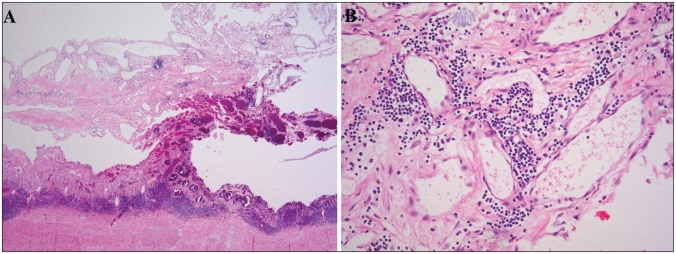
Fig. 2.
(A) Histological examination revealed that the tumour in the mesentery contained numerous dilated lymphatic spaces of varying sizes within collagenous stroma (hematoxylin and eosin stain, original magnification ×20). (B) Lymphatic spaces were lined by flattened endothelial cells, and small collections of lymphocytes were scattered throughout the lesion (hematoxylin and eosin stain, original magnification ×100).

