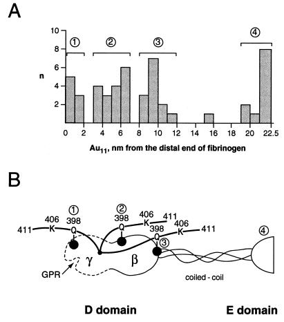
Figure 4.
A histogram showing the distribution of Au11 clusters on fibrinogen molecules (A) and an aligned diagram of a fibrinogen half-molecule illustrating the orientation of the C-terminal γ chain in the fibrinogen D domain (B). Gold clusters were distributed in four locations on the molecule: 1, distal D domain; 2, middle D domain; 3, proximal D domain; and 4, E domain. The gold clusters (•) that label the γ chains at γ398Q or γ399Q are designated simply as “398.”

