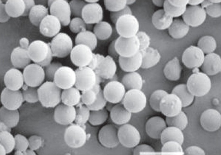
Fig. 1.

Scanning Electron Microphotograph of pentoxifylline loaded Poly(ε-caprolactone) microspheres
Scanning electron microphotograph of pentoxifylline loaded Poly(ε- caprolactone) microspheres was recorded at 200 X magnification to characterize shape and surface properties of the microspheres.
