Figure.
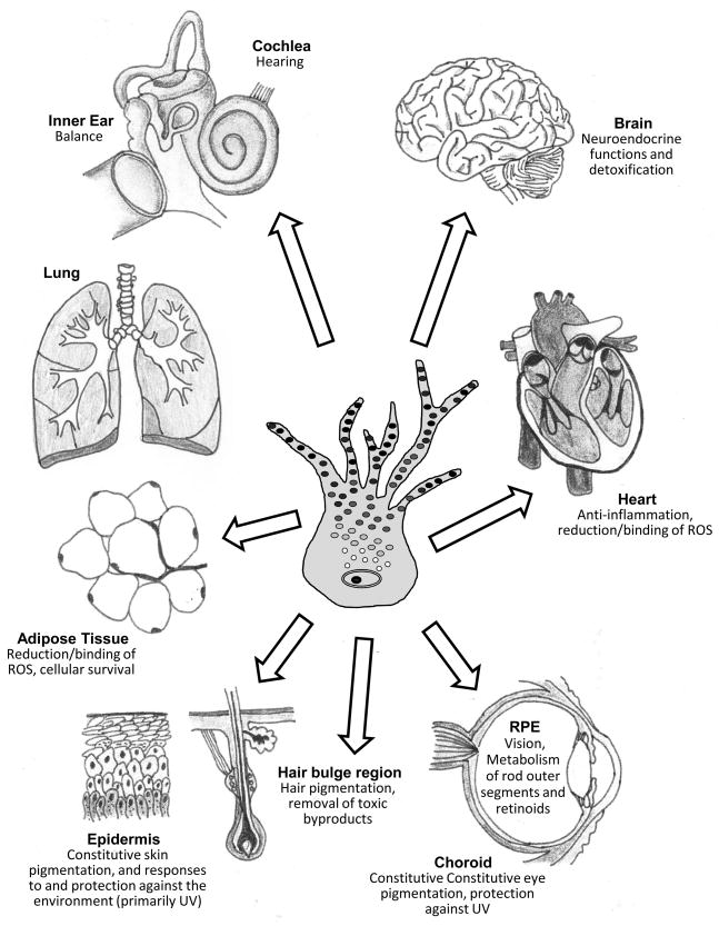
Schematic showing the known distribution of melanocytes in various human tissues, including the skin (epidermis and hair bulbs, bottom left), adipose tissue (lower left), lung (upper left), ear (inner ear and cochlea, top left), brain (top right), heart (right) and eye (retinal pigment epithelium and choroid, bottom right).

