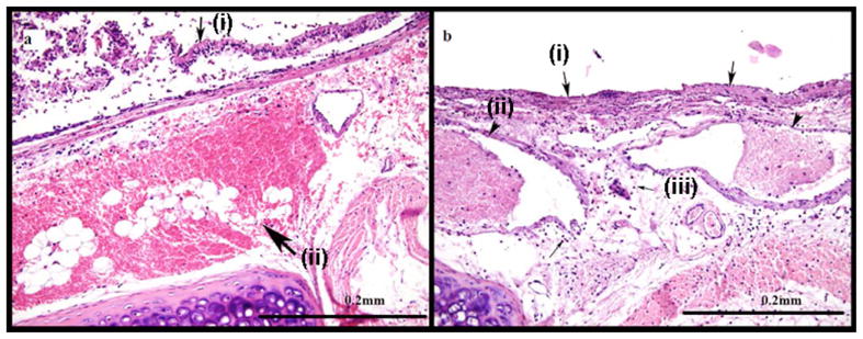
Fig. 3.

Images from an animal with mustard exposure. (a) Intraepithelial blisters [small arrow (i)] and extensive hemorrhage in the wall of the trachea [large arrow (ii)] are visible using HE staining after exposure to CEES. (b) Diffuse mucosal ulceration [large arrows (i)], vascular thrombi [arrow heads (ii)] and inflammation [small arrows (iii)] are also visible in the histology sample.
