Figure 4.
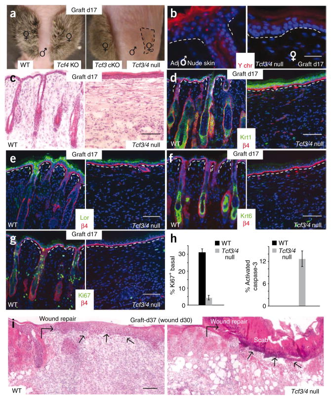
Skin grafting permits evaluation of the long-term consequences of Tcf3 and Tcf4 ablation in skin. (a) After 17 d of engraftment of female skins onto the backs of male nude mice, Tcf3 cKO and Tcf4 KO skins are indistinguishable from the wild type, whereas Tcf3/4-null grafts (area indicated by dashed line) shrink and show no hair. (b) Y-chromosome FISH shows that the engrafted Tcf3/4-null epidermis is still female and is not derived from the male nude epidermis surrounding the graft. (c) H&E staining reveals absence of hair follicles from Tcf3/4-null skin grafts. (d–g) Differentiation of the epidermis still occurs in Tcf3/4-null skin, as shown by immunolabeling for Krt1 (spinous) and loricrin (granular) markers. (f) Although epidermis of Tcf3/4-null grafts appears thicker, no immunolabeling was detected for Krt6, which marks the hair follicle ORS (companion layer) of normal skin and the suprabasal epidermis of hyperproliferative skin. (g) Ki67 immunolabeling reveals a paucity of proliferative keratinocytes within day 17 Tcf3/4-null skin grafts. (h) Quantification of Ki67 and activated caspase 3 staining. Error bars indicate s.d. (i) Tcf3/4-null engrafted skin is defective in reepithelialization after wounding at day 30 after engraftment. Shown are H&E-stained skins 7 d after wounding. Arrows indicate sites where reepithelialization has occurred to repair the wound, in control but not in Tcf3/4-null skin. Scale bars indicate 20 μm in b and 100 μm in c–g and i.

