Figure 6.
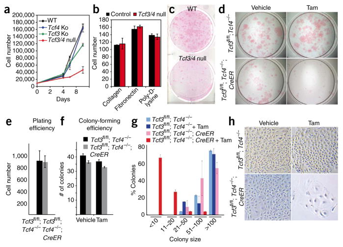
Loss of Tcf3 and Tcf4 results in inability of cultured epidermal keratinocytes to undergo long-term self-renewal. (a) Tcf3/4-null primary mouse keratinocyte cultures show a growth defect. (b) No differences in cell-substratum adhesion are seen when equal numbers of primary mouse keratinocytes are plated onto various matrices and quantified after 1 h. (c) Rhodamine B staining of primary mouse keratinocytes cultured for 10 d reveals a growth defect in the absence of Tcf3 and Tcf4. (d–h) Inducible deletion of Tcf3 from Tcf4−/− cells affects their growth potential. Cultured primary mouse keratinocytes from epidermis of Tcf3fl/fl; Tcf4−/− P0 mice were passaged twice and then infected with retrovirus expressing either green fluorescent protein (GFP) alone or GFP with tamoxifen-inducible Cre (transgene cre-Esr1; referred to here as ‘CreER’). Infected cells were isolated by flow cytometry based on GFP level, and 1 d after plating, cells were treated with tamoxifen or vehicle control to induce Cre recombinase activity. Twelve days after plating, cells were fixed and stained with rhodamine B to visualize and quantify the numbers and sizes of colonies (those with at least four cells, 72 h after plating). Plating (e,f) and colony-forming (f) efficiencies were comparable between wild-type and Tcf3/4-null cells, but the average size of Tcf3/4-null colonies was markedly smaller than that of wild-type colonies (g) and the morphology of the mutant cells revealed signs of premature differentiation (h). All error bars indicate s.d.

