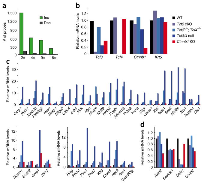
Figure 8.
Ablation of Tcf3 and Tcf4 in skin leads to an upregulation of gene expression. (a) Basal epidermal keratinocytes were purified by flow cytometry from E17.5 skins of Tcf3/4-null and wild-type mice, and their mRNAs were subjected to microarray analyses as outlined in Online Methods. Comparative analysis scored genes as being increased (‘Inc’) or decreased (‘Dec’) by the fold indicated. Most genes scored as upregulated upon loss of Tcf3 and Tcf4. (b–d) Real-time PCR analyses of mRNAs from flow cytometry–purified basal epidermal keratinocytes of wild-type, Tcf3 cKO, Tcf3+/−; Tcf4−/−, Tcf3/4-null and Ctnnb1 cKO E17.5 embryos. Genes chosen for analyses in b are controls to verify the status of Tcf3, Tcf4 and Ctnnb1 relative to comparable levels of the basal marker Krt5. Genes chosen for analyses in c scored as upregulated in Tcf3/4-null compared to wild-type epidermis and are known to directly bind to Tcf3 in embryonic stem cells20. Genes upregulated in E17.5 Tcf3/4-null basal epidermis were mostly unchanged (top) in Ctnnb1-null basal cells. A minority were either slightly increased or decreased (bottom panels). (d) A few genes scoring as decreased in the Tcf3/4-null samples also show reductions in expression when β-catenin is lost. Some of these are Wnt target genes, including Dkkl1 and Sostdc1.

