
Figure 1.
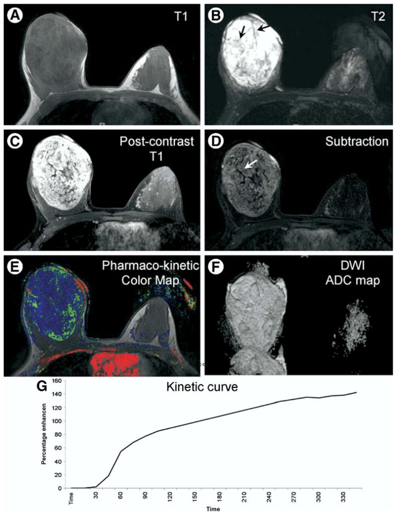
A 25 year old Caucasian female presented with a painless enlarging right breast mass. (A) Axial T1 weighted image shows a markedly enlarged right breast with a large oval well defined low signal mass. (B) Axial T2 weighted image with fat suppression shows the mass exhibiting bright T2 signal and dark internal septations (black arrows). (C, D) Axial post-contrast T1 weighted image with fat suppression and subtraction image show a heterogeneous enhancement pattern of the mass with non-enhancing septations (white arrow). (E) Axial pharmaco-kinetic post-processing color map shows the lesion with blue and green colors denoting low permeability and Kep values. (F) Axial ADC map shows th mass has high signal denoting high ADC value. (G) The corresponding kinetic curve of the lesion shows an enhancement pattern of the persistently enhancing (type 1a) type.