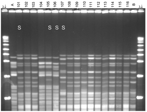
Figure 1.
Pulsed-field gel electrophoresis (PFGE) analysis of chromosomal DNA from pharyngeal meningococcus isolates (stained with ethidium bromide). Whole chromosome DNA macrorestriction fragments were generated by digestion with endonuclease SpeI. Shown are examples of sequence type (ST) prediction by PFGE in carried meningococci and diversity among STs. S, isolates tested by multilocus sequence typing (MLST). Lanes λ (arrows), PFGE marker I (Boehringer Mannheim, Mannheim, Germany); lane A, ST-2881, meningitis case isolate, Niger 2003; lanes 101, 102, and 103, ST-11, W135:2a:P1.5,2; lanes 104 and 105, ST-2881, W135:NT:P1.5,2; lane 106, ST-4151, W135:NT:P1.5,2; lane 107, ST-112000, W135:NT:P1.5,2; lanes 108–116, ST-11, W135:2a:P1.5,2; lane B, meningitis case isolate, ST-11, Niger 2003. Isolates 101 and 107 were identified as ST-11 by MLST. Isolates 102, 103, 108, 109, and 111–116 are indistinguishable from isolate 101 and are therefore considered ST-11. The 19 ST-11 isolates had 4 different PFGE patterns, of which 3 are represented by isolates 101, 107, and 110. The pattern of isolate 107 is indistinguishable from the 2000 Hajj epidemic strain (not shown).

