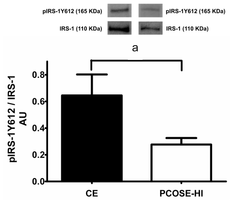
Figure 2.
Ratio between pIRS-1Y612 and IRS-1 in proliferative endometria from CE and PCOSE-HI women, assessed by Western blotting. Equal amounts of protein were loaded in each lane, and pIRS-1Y612 and IRS-1 band intensities were quantified by scanning densitometry and normalized to intensities observed for β-actin as internal control. A representative image of the media of bands obtained from 7 CE and 7 PCOSE-HI endometrial is shown. The results are expressed as AU and the values shown are mean ± SEM in CE and PCOSE-HI. a = P < 0.05 in PCOSE-HI compared with CE.

