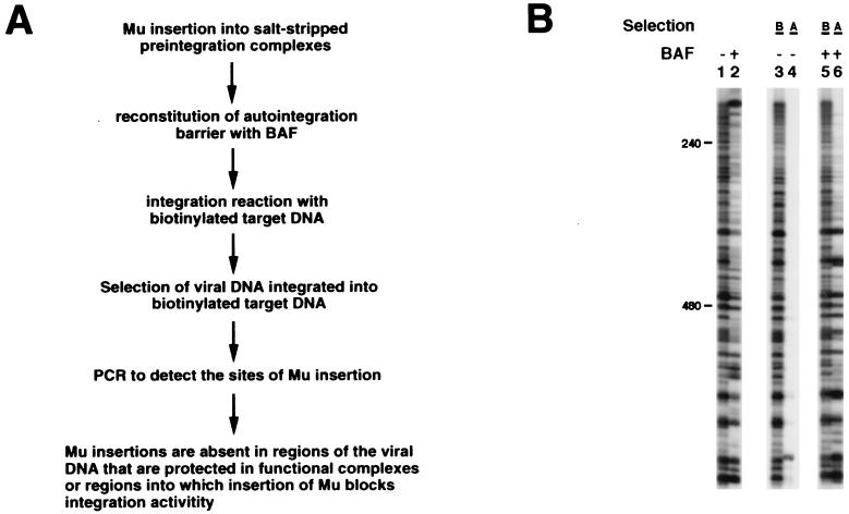
Figure 3.
Extensive regions at the ends of the viral DNA are required for integration activity. (A) Schematic depiction of the functional footprinting assay. (B) Functional footprints on the viral DNA U3 end. Lanes 1 and 2 are controls of standard MM-PCR footprinting with salt-stripped MLV preintegration complexes reconstituted with 0 or 2 ng BAF, respectively. Reconstitution reactions in the functional footprinting experiments contained 0 (lanes 3 and 4) or 20 ng BAF (lanes 5 and 6). DNA samples before and after biotin selection are marked with B and A, respectively. In the absence of reconstitution of the autointegration barrier with BAF, intermolecular integration was very inefficient, and therefore little signal is seen in lane 4.

