
Figure 3.
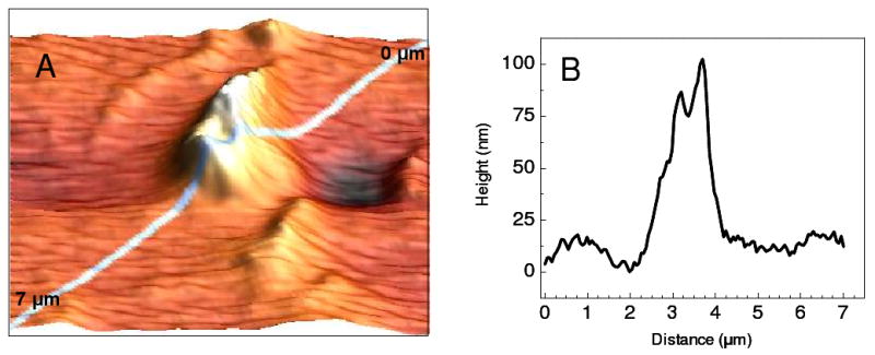
A 5 μm × 5 μm AFM image of single wall carbon nanotubes is depicted in A. The topographic profile obtained from the top right corner to the bottom left corner is shown in B.
Official websites use .gov
A
.gov website belongs to an official
government organization in the United States.
Secure .gov websites use HTTPS
A lock (
) or https:// means you've safely
connected to the .gov website. Share sensitive
information only on official, secure websites.
A 5 μm × 5 μm AFM image of single wall carbon nanotubes is depicted in A. The topographic profile obtained from the top right corner to the bottom left corner is shown in B.