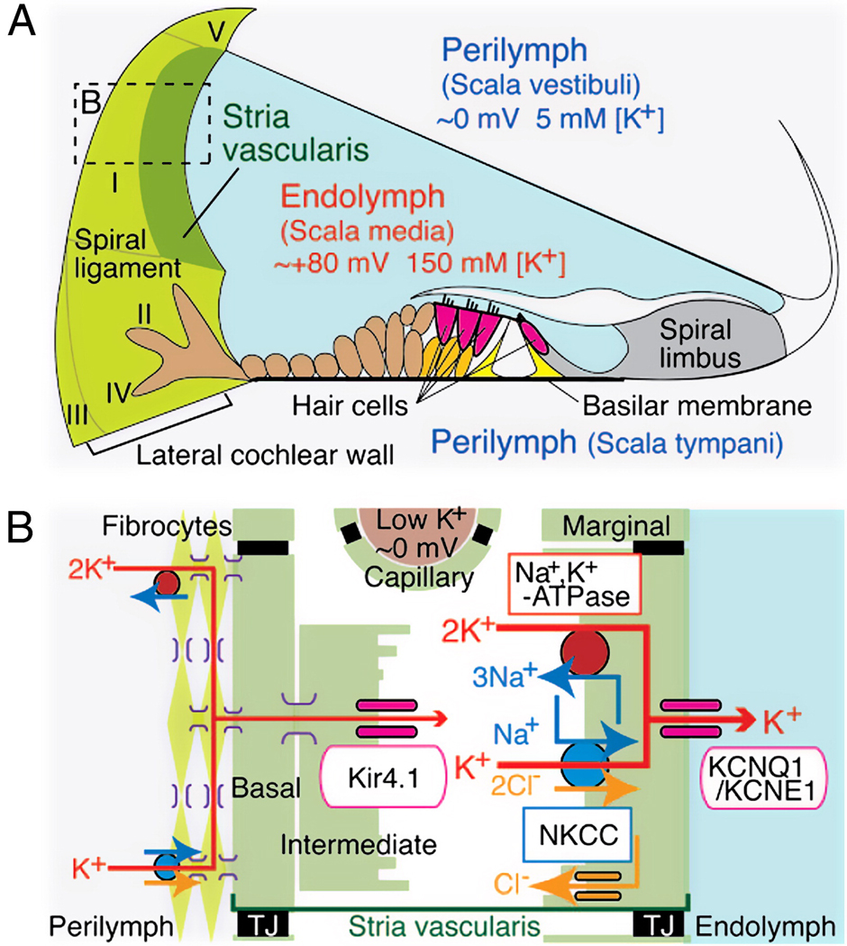
Figure 1.
Schematic structure of the cochlear duct with the lateral wall. A. Appropriate ionic composition of the endolymph and the EP require that an ion barrier line scala media, the intrastrial space, and strial capillaries. A nearly continuous cellular network guides K+ from the organ of Corti through finger-like root cells into (primarily) type II and I fibrocytes. Fibrocytes must take up K+ from the exracellular space around root cells. Locations of five major types of fibrocytes are indicated by roman numerals. B. Schematic enlargement of the boxed area in A depicts the cells and components required to generate the EP. Type I fibrocytes, strial basal cells, intermediate cells, as well as capillary pericytes (not shown) are joined by gap junctions composed mostly of connexins 26 and 30. K+ enters the intrastrial space through Kir4.1, then through marginal cells via Na+/K+-ATPase, the NKCC ion exchanger, and KCNQ1/KCNE1 channel complexes. TJ, tight junction. (Reprinted with permission from (Nin et al., 2008))

