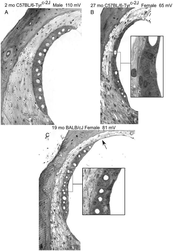
Figure 4.
Examples of lateral wall in the lower cochlear base of young B6 albino (A), old B6 albino (B), and old BALB/c (C). Age, gender, and basal turn EP are indicated. Aging was associated with strial thinning and marginal cell loss in both strains. Severity of capillary loss and ligament thinning in B are atypical for this strain. BALB (C) features greater loss of marginal cells along the luminal surface. Marginal cells in C inset show dense staining and retraction of processes. Arrow in C denotes somewhat unusual strial atrophy.

