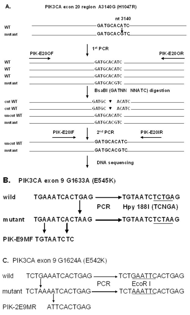
Fig. 1. The schematic of the mutant-enriched sequencing methods for detecting PIK3CA mutations, H1047R, E545K and E542K.
A. The protocol for detectingPIK3CA mutation H1047R. Enzyme BsaBI specifically cuts the wild-type sequences of the exon 20, but not the mutant copies with A3140G nucleotide alteration. After digesting the first PCR product with the enzyme BsaBI, the second PCR selectively amplifies the mutant copies. B. A unique restriction enzyme site Hpy188I was introduced by mismatch PCR for detecting PIK3CA mutation E545K. The mismatch primer (PIK-E9MF) has two A→ T nucleotide substitutions in the forward primer to create a unique enzyme site Hpy188I in the wild-type sequences of the PIK3CA exon 9, but not the mutant sequences. C. The mismatch primer PIK-2E9MR was designed to create a unique restriction enzyme site EcoRI for enriching PIK3CA mutation E542K (G1624A) and E542G (A1625G) with the similar strategy for the hot-spot mutation E545K.

