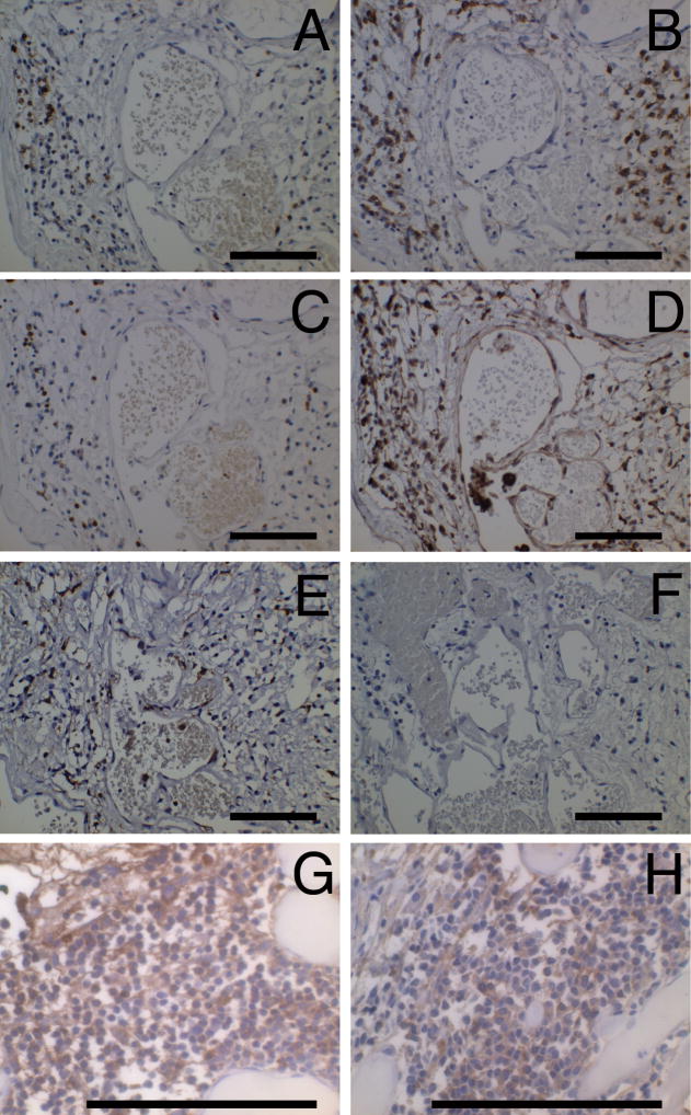
Figure 1.
Positive immune cells in CCM lesions. (A) B cells (CD20), (B) plasma cells (CD138), (C) T cells (CD3), (D) monocytes/macrophages (CD68), (E) antigen presenting cells (HLA-DR) and (F) negative control from the same area of the same specimen. (G) IgG positive cells and (H) IgM positive cells from two other specimens. Original magnification is 50 X (A–F) and 132 X (G, H). Scale bars are 100 μm.

