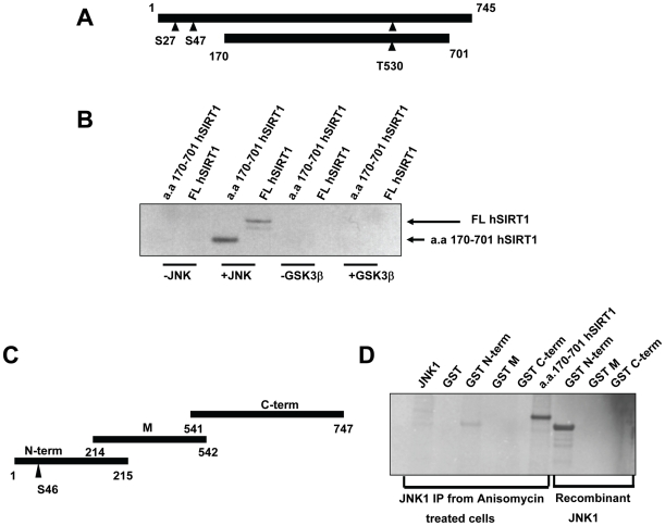
Figure 4. SIRT1 is phosphorylated by JNK1.
A, Schematic representation of full length human SIRT1 (FL hSIRT1) protein and a truncated form (aa 170–701 hSIRT1). Triangles show putative JNK1 phosphorylation sites. B, phosphorylation of SIRT1 by recombinant JNK1. FL hSIRT1 or a.a. 170–701 hSIRT1 was incubated with recombinant JNK1 or GSK3β in the presence of γ-[32P]-ATP. C, Schematic representation of mouse SIRT1 GST fragments. Triangle represents the putative JNK phosphorylation site. D, In vitro phosphorylation of mouse SIRT1 GST when treated with JNK1 immunoprecipitates from anisomycin-treated C2C12 cells or recombinant JNK1. Truncated hSIRT1 (a.a. 170–701 hSIRT1) was used as positive control.

