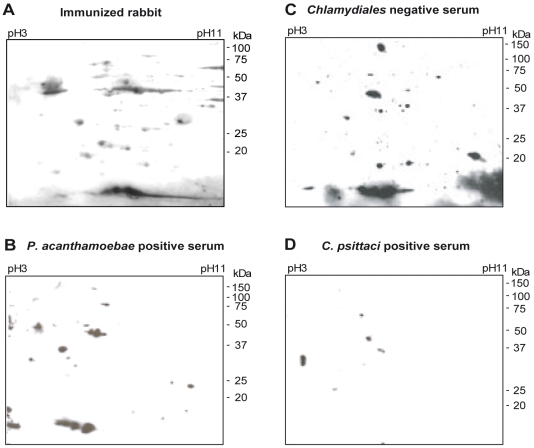
Figure 1. 2D patterns of the immunoreactive proteins of P. acanthamoebae.
Proteins of P. acanthamoebae separated by 2D gel electrophoresis were probed with (A) serum from immunized rabbit #1, (B) a Chlamydiales negative human serum, (C) a P. acanthamoebae positive human serum, and, (D) a C. psittaci positive human serum. Five immunogenic proteins are numbered in reference to the following figures.

