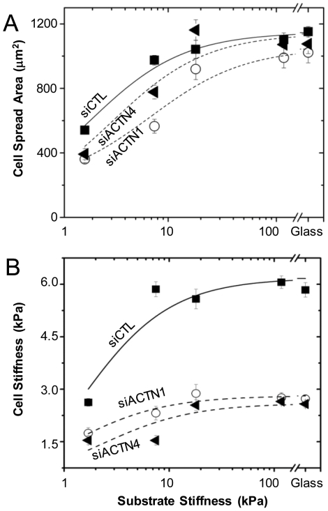
Figure 3. Contributions of α-actinin isoforms to cell-ECM rigidity sensing.
(A) Projected cell-ECM adhesion area of siCTL (squares), siACTN1 (circles), and siACTN4 (triangles) cells on collagen I-coated polyacrylamide ECMs of varying elasticity. Cell spreading differences between control and α-actinin depleted cells are statistically significant (p<0.001) for both isoforms on 2 kPa and 8 kPa ECMs. (B) Cortical cell elasticity of siCTL, siACTN1, and siACTN4 cells on variable-rigidity ECMs by AFM. Differences in cortical elasticity between control and α-actinin-depleted cells are statistically significant (p<0.001) for all ECM stiffness.

