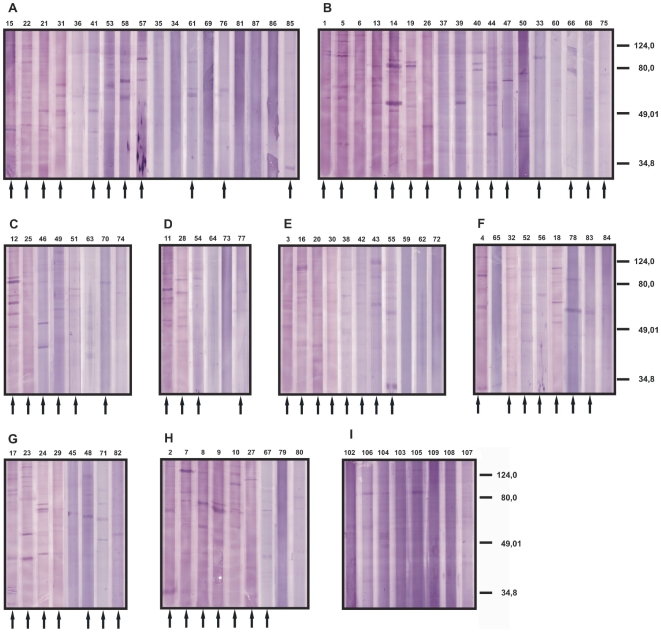
Figure 1. Pattern of seroreactivities of cutaneous lymphoma patients against the mycosis fungoides cell line MyLa.
Total protein extract of the tumor cells were separated by SDS-PAGE, blotted onto nitrocellulose and probed with the sera of the patients or of healthy control donors. The patients were diagnosed for mycosis fungoides (A male, B female), lymphomatoid papulosis (C), pleomorphic cutaneous T cell lymphoma (D), follicle center cell lymphoma (E), other cutaneous T cell lymphoma (F): CD30+ large cell lymphoma (sera 4, 65 and 84), cytotoxic cutaneous T cell lymphomas (sera 32 and 52), small cell to medium size cell T cell lymphoma (serum 56), CD8+ epidermotropic cytotoxic cutaneous T cell lymphoma (serum 78), parapsoriasis (serum 83) and Sezary syndrome (serum) 18) and other B cell lymphomas (G): diffuse large cell B cell lymphoma (sera 17 and 48), large cell B cell lymphoma (serum 24), mantle cell lymphoma (sera 23, 45 and 71), leukemia (serum 82) and one not specified B cell lymphoma (serum 29), not specified cutaneous lymphomas (H). The lanes marked with an arrow were rated as seropositive. As controls, sera of healthy donors were used (I). The numbers atop of each lane represent the numbers of the sera used and correspond to the patient numbering in Table S1. This numbering is used throughout this report.

