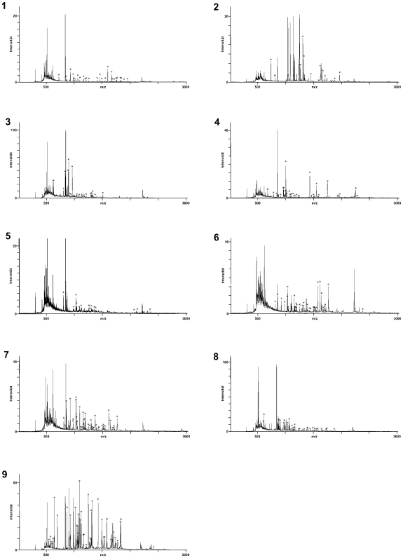
Figure 5. Identification of tumor-associated antigen defined by autologous sera.
The protein spots assigned to antigens were picked and treated with trypsin. The peptide mass fingerprints of the resulting protein fragments were determined by mass spectrometry. Twenty-two of the assigned antigens were identified as aconitase (spot and spectrum 1), β-tubulin (spot and spectrum 2), coronin (spot and spectrum 3), glutamate dehydrogenase (spot and spectrum 4), keratin 16 (spot and spectrum 5), lamin A (spot and spectrum 6), lamin C (spot and spectrum 7), lamin B1 (spots and spectrum 8) and vimentin (spots and spectrum 9). Lamin B1 was identified in two and vimentin in 13 different spots. Only one representative spectrum is shown each. The mass peaks marked with an asterisk correspond to peptide masses which were matched to the theoretical spectrum of the identified protein. The numbers of the spectra corresponds to the numbers of the spots in Figure 4.

