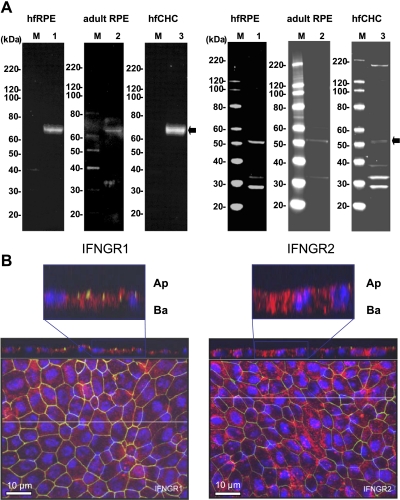
Fig. 1.
Expression and localization of IFNγ receptors. A: immunoblots of IFNγ receptor subunits 1 and 2 (IFNGR1 and IFNGR2) in human fetal and adult retinal pigment epithelium (RPE) and fetal choroidal (hfCHC) cells. M, molecular weight marker lanes; lane 1, primary hfRPE cell culture; lane 2, native human adult RPE; lane 3, primary hfCHC cells. B: immunofluorescence localization of IFNγ receptor in hfRPE cells. For IFNGR1 and IFNGR2, cross section through the z plane is shown at top. In each case, x-y plane is shown as an en face view of the apical membrane (maximum-intensity projection through the z-axis). ZO-1 (green) stains tight junctions; 4,6-diamidino-2-phenylindole (DAPI, blue) labels nuclei. Inset at higher gain shows a z-section above each panel; note that IFNGR1 and IFNGR2 are mainly located on the basolateral membrane (Ba). Ap, apical membrane.

