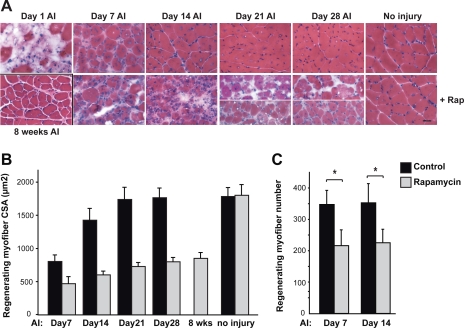
Fig. 1.
Muscle regeneration is impaired by rapamycin (Rap). Injury of the hindlimb tibialis anterior (TA) muscle was induced by BaCl2 injection in 10-wk-old male mice. The “no injury” control muscles received saline injection. Where indicated, Rap (1 mg/kg) was administered daily via intraperitoneal injection starting on day 1 after injury (AI). Vehicle was injected in all mice not receiving Rap. On various days AI, the animals were euthanized and TA muscles were isolated and cryosectioned. A: TA muscle cross-sections were stained with hematoxylin and eosin (H&E). Scale bar represents 50 μm. Two representative images are shown for day 21 and day 28 to demonstrate the abnormal muscle structure as well as smaller regenerating myofibers. B: cross-sectional area (CSAs) of regenerating myofibers, identified by their central nuclei on H&E-stained sections, were quantified. At least 100 regenerating myofibers were measured for each muscle section. C: number of regenerating myofibers was counted in an area of 614,400 μm2 for each sample. For all the data, the average results (n = 7 mice for each data point) are shown, with error bars representing SD. Paired two-tailed t-tests were performed to compare data from control and rapamycin-treated samples at each time point in C. *P < 0.01.

