
Figure 1. Amino acid frequencies in minor grooves.
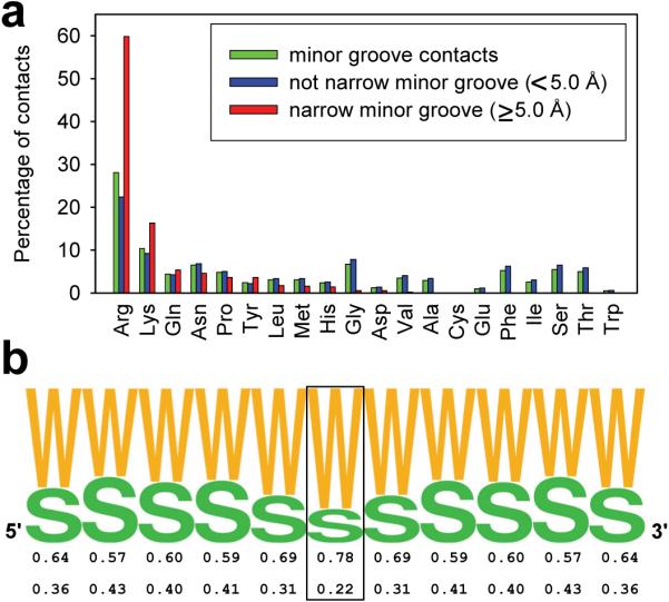
(a) Histograms for each amino acid illustrate the frequency with which they are observed in any minor groove (green), in minor grooves with a width of ≥5.0 Å (blue), and in narrow minor grooves of <5.0 Å width (red). (b) Frequency of AT (W) and GC (S) base pairs in sequences of 229 sites contacted by arginines in narrow minor grooves. The central base pair (boxed) is contacted by arginine. Frequencies are symmetrized by using both complementary strands.